
Chronic Obstructive Pulmonary Disease- COPD
What It Is, The Causes and Detection
Part l
Chronic Obstructive Pulmonary Disease- What Is It?
Chronic obstructive pulmonary disease (COPD), also called chronic obstructive lung disease, is a term that is used for two closely related diseases of the respiratory system: chronic bronchitis and emphysema. In many patients these diseases occur together, although there may be more symptoms of one than the other. Most patients with these diseases have a long history of heavy cigarette smoking.
COPD gets gradually worse over time. At first there may be only a mild shortness of breath and occasional coughing. Then a chronic cough develops with clear, colorless sputum. As the disease progresses, the cough becomes more frequent and more and more effort is needed to get air into and out of the lungs. In later stages of the disease, the heart may be affected. Eventually death occurs when the function of the lungs and heart is no longer adequate to deliver oxygen to the body’s organs and tissues.
Cigarette smoking is the most important risk factor for COPD; it would probably be a minor health problem if people did not smoke. Other risk factors include age, heredity, exposure to air pollution at work and in the environment, and a history of childhood respiratory infections. Living in low socioeconomic conditions also seems to be a contributing factor.
It has been estimated that six and a half million people have been diagnosed with some form of COPD and over 15 million more have been left undiagnosed.. It is the fourth leading cause of death in the United States. Between 1980 and 1990, the total death rate from COPD increased by 22 percent. In 1990, it was estimated that there were 84,000 deaths due to COPD, approximately 34 per 100,000 people. New government data based on a 1998 prevalence survey suggest that three million Americans have been diagnosed with emphysema and nine million were affected by chronic bronchitis .Although COPD is still much more common in men than women, the greatest increase in the COPD death rate between 1979 and 1989 occurred in females, particularly in black females (117.6 percent for black females vs. 93 percent for white females). These increases reflect the increased number of women who smoke cigarettes.
COPD attacks people at the height of their productive years, disabling them with constant shortness of breath. It destroys their ability to earn a living, causes frequent use of the health care system, and disrupts the lives of the victims’ family members for as long as 20 years before death occurs.
In 1990, COPD was the cause of approximately 16.2 million office visits to doctors and 1.9 million hospital days. The economic costs of this disease are enormous. In 1989, an estimated $7 billion was spent for care of persons with COPD and another $8 billion was lost to the economy by lost productivity due to morbidity and mortality from COPD.
What Are Chronic Bronchitis and Emphysema?
Chronic bronchitis, one of the two major diseases of the lung grouped under COPD, is diagnosed when a patient has excessive airway mucus secretion leading to a persistent, productive cough. An individual is considered to have chronic bronchitis if cough and sputum are present on most days for a minimum of 3 months for at least 2 successive years or for 6 months during 1 year. In chronic bronchitis, there also may be narrowing of the large and small airways making it more difficult to move air in and out of the lungs. begins in the smaller airways within the lungs and gradually advances to larger airways. It increases mucus in the airways and increases bacterial infections in the bronchial tubes, which, in turn, impedes airflow An estimated 12.1 million Americans have chronic bronchitis.
In emphysema there is permanent destruction of the alveoli, the tiny elastic air sacs of the lung, because of irreversible destruction of a protein in the lung called elastin that is important for maintaining the strength of the alveolar walls. The loss of elastin also causes collapse or narrowing of the smallest air passages, called bronchioles, which in turn limits airflow out of the lung.
In the general population, emphysema usually develops in older individuals with a long smoking history. However, there is also a form of emphysema that runs in families. People with familial emphysema have a hereditary deficiency of a blood component, alpha-l-protease inhibitor, also called alpha-l-antitrypsin (AAT). The number of Americans with this genetic deficiency is quite small, probably no more than 70,000. It is estimated that 1 in 3,000 newborns have a genetic deficiency of AAT, and 1 to 3 percent of all cases of emphysema are due to AAT deficiency.
The destruction of elastin that occurs in emphysema is believed to result from an imbalance between two proteins in the lung–an enzyme called elastase, which breaks down elastin, and AAT, which inhibits elastase. In the normal individual, there is enough AAT to protect elastin so that abnormal elastin destruction does not occur. However, when there is a genetic deficiency of AAT, the activity of the elastase is not inhibited and elastin degradation occurs unchecked. If individuals with a severe genetic deficiency of alpha-l-protease inhibitor smoke, they usually have symptoms of COPD by the time they reach early middle age. Deficiency of alpha-l-protease inhibitor can be detected by blood tests available through hospital laboratories. People from families in which relatives have developed emphysema in their thirties and forties should be tested for AAT deficiency. If a deficiency is found, it is critical for these people not to smoke.
Some scientists believe that nonfamilial emphysema, usually called “smoker’s emphysema,” also results from an imbalance between elastin-degrading enzymes and their inhibitors. The elastase-AAT imbalance is thought to be a result of the effects of smoking, rather than inherited as in familial emphysema. Some evidence for this theory comes from studies on the effect of tobacco smoke on lung cells. These studies showed that tobacco smoke stimulates excess release of elastase from cells normally found in the lung. The inhaled smoke also stimulates more elastase-producing cells to migrate to the lung, which in turn causes the release of even more elastase. To make matters worse, oxidants found in cigarette smoke inactivate a significant portion of the elastase inhibitors that are present, thereby decreasing the amount of active antielastase available for protecting the lung and further upsetting the elastase-antielastase balance.
Scientists believe that, in addition to smoking-related processes, there must be other factors that cause emphysema in the general population since only 15 to 20 percent of smokers develop emphysema. The nature and role of these other factors in smokers’ emphysema are not yet clear.
What Are the Risk Factors for COPD?
Long-term smoking is the most frequent cause of COPD. It accounts for 80 to 90 percent of all cases. A smoker is 10 times more likely than a nonsmoker to die of COPD.
Other risk factors include:
- Heredity
- Secondhand smoke
- Exposure to air pollution at work and in the environment
- A history of childhood respiratory infections
What Goes Wrong With the Lungs and Other Organs in Chronic Obstructive Pulmonary Disease?
The most important job that the lungs perform is to provide the body with oxygen and to remove carbon dioxide. This process is called gas exchange, and the normal anatomy of the lungs serves this purpose well. The lungs contain 300 million alveoli whose ultrathin walls form the gas exchange surface. Enmeshed in the wall of each of these air sacs is a network of tiny blood vessels, the capillaries, which bring blood to the gas exchange surface. When a person inhales, air flows from the nose and mouth through large and small airways into the alveoli. Oxygen from this air then passes through the thin walls of the inflated alveoli and is taken up by the red blood cells for delivery to the rest of the body. At the same time, carbon dioxide leaves the blood and passes through the alveolar walls into the alveoli. During exhalation, the lung pushes the used air out of the alveoli and through the air passages until it escapes from the nose or mouth.
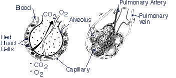
Gas Exchange
Inhaled air travels through the airways to the alveoli. Blood is pumped out of the heart through the pulmonary arteries to a network of capillaries that surround the alveoli. The oxygen of the inhaled air diffuses out of the alveoli into the blood while carbon dioxide in the blood moves into the alveoli to be exhaled. The oxygen-rich blood is returned to the heart through the pulmonary veins.
When COPD develops, the walls of the small airways and alveoli lose their elasticity. The airway walls thicken, closing off some of the smaller air passages and narrowing larger ones. The passageways also become plugged with mucus. Air continues to get into alveoli when the lung expands during inhalation, but it is often unable to escape during exhalation because the air passages tend to collapse during exhalation, trapping the “stale” air in the lungs. These abnormalities create two serious problems which affect gas exchange:
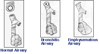
Air Ways
- Blood flow and air flow to the walls of the alveoli where gas exchange takes place are uneven or mismatched. In some alveoli there is adequate blood flow but little air, while in others there is a good supply of fresh air but not enough blood flow. When this occurs, fresh air cannot reach areas where there is good blood flow and oxygen cannot enter the bloodstream in normal quantities.
- Pushing the air through narrowed obstructed airways becomes harder and harder. This tires the respiratory muscles so that they are unable to get enough air to the alveoli. The critical step for removing carbon dioxide from the blood is adequate alveolar airflow. If airflow to the alveoli is insufficient, carbon dioxide builds up in the blood and blood oxygen diminishes. Inadequate supply of fresh air to the alveoli is called hypoventilation. Breathing oxygen can often correct the blood oxygen levels, but this does not help remove carbon dioxide. When carbon dioxide accumulation becomes a severe problem, mechanical breathing machines called respirators, or ventilators, must be used.
Pulmonary function studies of large groups of people show that lung function–the ability to move air into and out of the lungs–declines slowly with age even in healthy nonsmokers. Because healthy nonsmokers have excess lung capacity, this gradual loss of function does not lead to any symptoms. In smokers, however, lung function tends to worsen much more rapidly. If a smoker stops smoking before serious COPD develops, the rate at which lung function declines returns to almost normal. Unfortunately, because some lung damage cannot be reversed, pulmonary function is unlikely to return completely to normal.
COPD also makes the heart work much harder, especially the main chamber on the right side (right ventricle), which is responsible for pumping blood into the lungs. As COPD progresses, the amount of oxygen in the blood decreases which causes blood vessels in the lung to constrict. At the same time many of the small blood vessels in the lung have been damaged or destroyed as a result of the disease process. More and more work is required from the right ventricle to force blood through the remaining narrowed vessels. To perform this task, the right ventricle enlarges and thickens. When this occurs the normal rhythm of the heart may be disturbed by abnormal beats. This condition, in which the heart is enlarged because of lung problems, is called cor pulmonale. Patients with cor pulmonale tire easily and have chest pains and palpitations. If an additional strain is placed on the lungs and heart by a normally minor illness such as a cold, the heart may be unable to pump enough blood to meet the needs of other organs. This results in the inability of the liver and kidneys to carry out their normal functions, which leads to swelling of the abdomen, legs, and ankles.
Another adjustment the body makes to inadequate blood oxygen is called secondary polycythemia, an increased production of oxygen-carrying red blood cells. The larger than normal number of red blood cells is helpful up to a point; however, a large overpopulation of red cells thickens the blood so much that it clogs small blood vessels causing a new set of problems. People who have poor supply of oxygen usually have a bluish tinge to their skin, lips, and nailbeds, a condition called cyanosis.
Too little oxygen and too much carbon dioxide in the blood also affect the nervous system, especially the brain, and can cause a variety of problems including headache, inability to sleep, impaired mental ability, and irritability.
What Is the Course of Chronic Obstructive Pulmonary Disease?
Daily morning cough with clear sputum is the earliest symptom of COPD. During a cold or other acute respiratory tract infection, the coughing may be much more noticeable and the sputum often turns yellow or greenish. Periods of wheezing are likely to occur especially during or after colds or other respiratory tract infections. Shortness of breath on exertion develops later and progressively becomes more pronounced with severe episodes of breathlessness (dyspnea) occurring after even modest activity.
A typical course of COPD might proceed as follows. For a period of about 10 years after cigarette smoking begins, symptoms are usually not very noticeable. After this, the patient generally starts developing a chronic cough with the production of a small amount of sputum. It is unusual to develop shortness of breath during exertion below the age of 40, after which it becomes more common and may be well developed by the age of 50. However, although all COPD patients have these symptoms, not all cigarette smokers develop a notable cough and sputum production, or shortness of breath.
Most patients with COPD have some degree of reversible airways obstruction. It is therefore likely that, at first, treatment will lead to some improvement or stability in lung function. But as COPD progresses, almost all signs and symptoms except cough and sputum production tend to show a gradual worsening. This trend can show fluctuations, but over the course of 4 or 5 years, a slow deterioration becomes evident.
Repeated bouts of increased cough and sputum production disable most patients and recovery from coughing attacks may take a long time. Patients with severe lung damage sleep in a semi-sitting position because they are unable to breathe when they lie down. They often complain that they awaken during the night feeling “choked-up,” and they need to sit up to cough.
Survival of patients with COPD is closely related to the level of their lung function when they are diagnosed and the rate at which they lose this function. Overall, the median survival is about 10 years for patients with COPD who have lost approximately two-thirds of their normally expected lung function at diagnosis.
The Global Initiative for Chronic Obstructive Lung Disease (GOLD) guidelines (April 2001) recommend the classification of disease severity into four stages 5:
Stage 0: At Risk—Chronic cough and sputum production. Lung function is normal, as measured by spirometry.5
Stage I: Mild COPD—Mild airflow limitation (FEV1/FVC < 70% but FEV1 > 80% predicted) and usually, but not always, chronic cough and sputum production. At this stage, the individual may not be aware that his or her lung function is abnormal.5
Stage II: Moderate COPD—Worsening airflow limitation (FEV1/FVC < 70%;30% < 80% predicted [IIA:50% < FEV1 < 80% predicted; IIB: 30% < FEV1 < 50% predicted]) and usually the progression of symptoms, with shortness of breath typically developing on exertion. This is the stage at which patients typically seek medical attention because of dyspnea or an exacerbation of their disease. The division into stages IIA and IIB is based on the fact that exacerbations are especially seen in patients with FEV1 below 50% predicted. The presence of repeated exacerbations has an impact on the quality of life of patients and requires appropriate management.5
Stage III: Severe COPD—Severe airflow limitation (FEV1/FVC < 70%;FEV1 < 30% predicted) or the presence of respiratory failure or clinical signs of right heart failure. Patients may have severe (Stage III) COPD, even if FEV1 is > 30% predicted, whenever these complications are present. At this stage, quality of life is appreciably impaired and exacerbations may be life-threatening.
How Is Chronic Obstructive Pulmonary Disease Detected?
Researchers are still looking for accurate methods to predict a person’s chances of developing airway obstruction. None of the current ways used to diagnose COPD detects the disease before irreversible lung damage occurs. While many measures of lung function have been developed, those most commonly used determine: 1) air-containing volume of the lung (lung volume), 2) the ability to move air into and out of the lung, 3) the rate at which gases diffuse between the lung and blood, and 4) blood levels of oxygen and carbon dioxide.
Lung volumes are measured by breathing into and out of a device called a spirometer. Some types of spirometers are very simple mechanical devices, which record volume changes as air is added to or removed from them. Other kinds are more sophisticated and use various types of electronic equipment to determine and record the volume of air moved into and out of the lungs. The three volume measures most relevant to COPD are forced vital capacity (FVC), residual volume (RV), and total lung capacity (TLC). The forced vital capacity is the maximum volume of air which can be forcibly expelled after inhaling as deeply as possible. Not all of the air in the lungs is removed when measuring the vital capacity. The amount remaining is called the residual volume. The total lung capacity is the combination of the forced vital capacity and residual volume. While most of the measured lung volumes or capacities change to some degree with COPD, residual volume usually increases quite markedly. This increase is the result of the weakened airways collapsing before all the normally expired air can leave the lungs. The increased residual volume makes breathing even more difficult and labored.
Because COPD results in narrowed air passages, a measure of the rate at which air can be expelled from the lungs can also be used to determine how severe the narrowing has become. In this test, the forced vital capacity maneuver, the patient is asked to inhale as deeply as possible, and on signal, exhale as completely and as rapidly as possible. The volume of air exhaled within 1 second is then measured. This value is referred to as the forced expiratory volume in 1 second (FEV1). When FEV1 is used as an indicator of lung function, the average rate of decline in patients with chronic obstructive lung disease is observed to be two to three times the normal rate of 20-30 milliliters per year. This volume may also be expressed in terms of the percent of the vital capacity, which can be expelled in 1 second. As COPD progresses, less air can be expelled in 1 second. A greater than expected annual fall in FEV1 is the most sensitive test for COPD and a fairly good predictor of disability and early death.
Another measure of lung function is called diffusing capacity. For this, a more complicated test determines the amount of gas, which can move in a given period of time from the alveolar side of the lung into the blood. A number of conditions can cause the diffusing capacity to decrease. However, in COPD the decrease is the result of the destruction of alveolar walls, which leads to a significant decrease in surface area for diffusion of oxygen into the blood.
Because the primary function of the lung is to remove carbon dioxide from the blood and add oxygen, another indicator of pulmonary function is the blood levels of oxygen and carbon dioxide. As chronic obstructive pulmonary disease progresses, the amount of oxygen in the blood decreases and carbon dioxide increases.
In most cases, it is necessary to compare the results of several different tests in order to make the correct diagnosis, and to repeat some tests at intervals to determine the rate of disease progression or improvement. Measurement of FEV1 and FEV1/FVC ratio should be a routine part of the physical examination of every COPD patient. It is hoped that current research will result in more accurate and earlier measures for detecting lung destruction and diminished function.
National Heart, Lung and Blood Institute, Division of Lung Diseases
